
2007 SPRING
MEETING of the MICHIGAN ASM |
STUDENT POSTERS (eligible for judging at the meeting)
Streptococcus iniae exploits the host environment to cause a
systemic infection
BETH A. LOWE* and MELODY N. NEELY
Wayne State University School of Medicine, Detroit, MI
Streptococcal systemic pathogens such as Streptococcus agalactiae and Streptococcus pneumoniae
cause serious morbidity and mortality worldwide. Although a great deal of valuable work
has been reported on streptococcal pathogenesis, achieving conclusive results on virulence
mechanisms has been problematic because many streptococcal species are human specific and
lack a good animal model. Our lab employs a zebrafish host model to study systemic
infections caused by S. iniae, a natural
systemic pathogen of both fish and humans. We have demonstrated that S. iniae systemic infections in zebrafish closely
mimic the clinical pathologies caused by S.
agalactiae and S. pneumoniae infections in humans. One virulence
factor found in common in systemic pathogens is polysaccharide capsule. The capsule is
important in circumventing the normal innate immune response of the host. In an S. iniae infection of zebrafish, the bacteria
quickly disseminate from the site of injection, the dorsal muscle, to all organs of the
fish. Host inflammatory cells aid in this process by engulfing and transporting the
bacteria from the muscle. We hypothesize that the capsule is protecting the bacteria in
this environment, and, that this mechanism requires a high level of regulation of capsule
expression. The specifics of regulation of capsule and its role in dissemination are
currently being investigated. The use of the zebrafish-S. iniae infection model will provide an
understanding of S. iniae pathogenesis, as well
as insight into the mechanisms used by streptococcal systemic pathogens to disseminate and
cause disease.
Characterization of NG1684, a cell contact regulated gene of Neisseria gonorrhoeae
Upon introduction to a new host it is essential that the exclusively human pathogen Neisseria gonorrhoeae adapt and respond to a changing host milieu, in which the initial interactions with host epithelial cells induce critical differential regulation of bacterial factors necessary for establishment of a successful infection. The primary goal of the research presented here is to examine NG1684, a gene that has been identified as being significantly upregulated in response to host epithelial cell contact. Transposon mutagenesis, quantitative real time PCR, and a tissue culture model of infection were employed to characterize the role of NG1684 in gonococcal infection and the regulation of NG1684 in response to host cell contact. We have determined that a transposon insertion in NG1684 in N. gonorrhoeae strain MS11 results in a decreased ability to invade the endometrial cell line Hec1B, although the ability of this mutant to adhere to these cells is indistinguishable from the wild type strain. Investigation of the flanking regions of NG1684 showed that this gene is divergently transcribed from farAB, which encodes an efflux pump responsible for conferring resistance to host derived antibacterial factors. The transcriptional regulator, FarR, binds to multiple sites in the intergenic region between NG1684 and farAB and has been shown to repress farAB expression. We have evidence to suggest that FarR also regulates NG1684 expression. FarR expression has been shown to be regulated by MtrR, which also regulates mtrCDE. This operon encodes a separate efflux pump responsible for resistance to a broad range of structurally diverse antimicrobial compounds. Moreover, mtrR expression was identified as being upregulated in response to host cell contact in addition to NG1684. This observation is consistent with our hypothesis that FarR represses NG1684, as an increase in MtrR would lead to a reduction of FarR and a subsequent increase in NG1684 expression. Based on the fact that FarR and MtrR mediate resistance to host derived antimicrobial agents in N. gonorrhoeae, we hypothesize that NG1684 may be involved in response to and/or resistance to antimicrobial compounds.
Streptococcus
pyogenes is an important human pathogen that can respond and adapt to changing
environments. Although in vitro analyses have
proven invaluable in determining specific streptococcal virulence factors, such studies
are limited in their ability to provide a natural environment in which the invading
pathogen responds directly, as well as indirectly, to the host conditions and defense
systems. The goal of this study is to identify S.
pyogenes virulence genes important for pathogenesis within the host using
signature-tagged mutagenesis. Currently there are no reports in the literature of this
technique being applied to S. pyogenes.
Moreover, a novel animal model, the zebrafish, is used, which has been proven to mimic the
responses involved in human streptococcal infection. The importance of this study is
underscored by the fact that the variety and severity of S. pyogenes infections are on the rise worldwide.
Thus, the current state of antimicrobial therapy is lacking in its aim to contain or
eradicate this seemingly ubiquitous human pathogen. Therefore, novel strategies are
necessary and can only be accomplished with the aid of in vivo analyses in determining the molecular
mechanisms involved in S. pyogenes pathogenesis.
Incidence of polyglutamine insertions (poly-Q) in Escherichia coli: A model for polyglutamine
expansion in genetic diseases in humans?
Background: Expansions of trinucleotide regions coding for glutamines (Qs) are characteristic of several human genetic diseases, including Huntington’s disease and cancer. The mechanisms mediating poly-Q expansions in human genetic diseases are not well understood. Recently, our lab discovered trinucleotide expansions coding for Qs in the tsr gene, the serine methyl-accepting chemotaxis receptor gene.The common laboratory E. coli strain, K-12, has a sequence coding for 4 Qs that could be the core of poly-Q expansion in E. coli. This study investigates the incidence of poly-Q expansions in the tsr gene of a subset of wild E. coli strains derived from different hosts.
Methods: Initially, expanded poly-Q strains were discovered during sequencing of the conserved region of the tsr gene in 20 diverse E. coli strains isolated from animal feces. Subsequently, PCR primers flanking the poly-Q expansion region were used to identify poly-Q regions of different lengths. Electrophoresis utilizing 3% agarose gels was used as a method to differentiate between the variable lengths of PCR products which are dependent upon tsr gene expansions. Searches of Genbank revealed additional tsr poly-Q variants.
Results: Survey of 37 wild E. coli strains revealed that the incidence of tsr gene expansions coding for poly-Q sequences of the same or longer length as K-12 is as follows: 4Q, 8 strains (22%); 7Q, 24 strains (65%); 13Q, 5 strains (14%). Presently, more strains are being surveyed; however, according to these results, most natural strains of E. coli have a sequence of 7 glutamines. Bioinformatic analysis revealed one 10Q strain (E. coli HS) and that O157:H7 and UTI pathogenic E. coli strains have 7Qs. Experiments are in progress to determine the stability of inheritance of the large poly-Q sequences.
Conclusion: E. coli has a naturally occurring variable poly-Q region. Studies to determine the mechanisms that bring about expansion or deletion of codons in this region may reveal mechanisms that mediate similar phenomena in human genetic diseases.
A study of common and
strain-specific Mycobacterium avium subspecies paratuberculosis infection induced transcriptome
changes in bovine
Mycobacterium
avium subspecies paratuberculosis (MAP) is
an intracellular pathogen that causes an economic burden to US dairy industries estimated
at over one billion dollars annually. MAP has
also been linked to some cases of human Crohn’s disease. A hallmark of MAP infection is survival in host
macrophages, cells that normally destroy ingested microbes.
As with other mycobacteria, survival in macrophages appears to be a key
determinant of pathogenesis associated with MAP infections.
Based on previous studies relating MAP infection with altered macrophage
gene expression, we hypothesized that different strains of MAP would have both common and
strain-specific effects on macrophage cell gene expression.
To test this hypothesis, we have now studied the effect of 10 different MAP
strains on macrophage gene expression profiles, with the ultimate goal of relating gene
expression differences to virulence and genetics of the MAP strains. Initial data analysis
suggests that there are over 120 macrophage genes whose expression is generally altered
following infection with any strain of MAP. Upon
hierarchical clustering using fold-change data, MAP strains isolated from different
species showed little initial host species similarity, but two highly virulent strains
clustered together, perhaps suggesting these two strains have similar effects on bovine
macrophage cells.
Oral Bacteria Counts
on Reused Water Bottles
Many people re-use water bottles after purchasing bottled water. This investigation was done in order to measure the numbers and types of bacteria found on the mouths of the bottles after repeated re-use. We found that even with washing, after multiple uses bacteria do accumulate on the mouths of the bottles. However, as expected, the bacteria identified were all normal resident oral flora.
Using PCR-DGGE to
determine the bacterial diversity and identity of cloacal samples from Tree Swallows (Tachycineta bicolor)
Cloacal swabs were collected from 36
nestling Tree Swallows (Tachchycineta bicolor),
DNA was extracted from the swabs and amplified. Amplified DNA was separated by use of
denaturing gradient gel electrophoresis (DGGE). Bands of interest were picked,
re-amplified, sequenced, and identified using GenBank. Gradient gel banding patterns were
analyzed using Gel2K and similarity indices were calculated using a Jaccard’s
Similarity Index. The similarity of bands separated by DGGE ranged drastically between
individuals within a nest (4.76 - 100%). These results indicate that in some cases nest
mates appear to have an identical cloacal microbial flora, whereas in other nests a
uniform microbial flora was not found. DNA sequencing of DGGE bands identified two known
avian pathogens present in a number of nestlings, Suttonella
ornithocola and Mycoplasma sturnidae. S. ornithocola was found in 10 of the 24 birds
sampled and M. sturnidae was found in 15 of the 24 birds sampled. Candida albicans is
a dimorphic (hyphal or yeast), opportunistic fungal pathogen, which poses a significant
clinical threat to immunocompromised individual. Diseases
associated with this fungus ranges from systemic to superficial mucosal hypersensitivity
responses. The mechanisms by which Candida persists at mucosal surfaces in the face
of an adaptive response are unclear. Candida produces immunomodulatory oxylipins
that cross-react functionally with host eicosanoids, which are considered to play
important roles in innate and adaptive immune responses.
At the mucosal surface, dendritic cells (DC) direct the type of T-cell
responses after interacting with pathogen at mucosal surface. Yeast forms induce
protective DC1/Th1 responses, while more virulent hyphal forms induce non-protective
DC2/Th2 responses. Interestingly, previous studies have also showed that host eicosanoid
(PGE2) increases the transformation from yeast-to-hyphae which can cause the
fungus to become persistent and virulent. Our objective is to characterize the role of
prostaglandins produced by the host and this fungus in pathogenesis in vivo and during Candida-dendritic
cell interactions. We hypothesize that production of oxylipin by both Candida and
host are required for persistent infection. We are testing this hypothesis by examining
effects of host and fungal prostaglandins on DC cytokine profiles and also maturation and
activation markers in the presence of yeast or hyphae. To address whether the effects of
prostaglandins are host-derived, we are testing responses in DCs isolated from COX-2
deficient mice (an enzyme required for synthesis of eicosanoids). Understanding the
mechanisms by which Candida modulates our immune system will provide new strategies
to treat infection caused by this pathogen.
Establishment of a Method to Examine Plant Pathogen
Effectors
Our aim is to create a model system to study the action of plant
pathogens when they infect plant cells. We have established an expression system in Saccharomyces cerevisiae to study HopM1. The
expression vector contains an inducible GAL1
promoter for the expression of HopM1. It also contains a V5 epitope that will be used for
visualization of HopM1 and 6xHIS tag for
purification. We have begun to amplify additional Hop genes using Polymerase Chain Reaction (PCR).
The fragments are first cloned into the pCR-Blunt II-TOPO vector and sequenced for
verification of correctness. Subsequently they are transferred to yeast plasmids for
expression. HopM1 when expressed in yeast is lethal on solid media. Examination of the
lethal effect using a titer assay has not duplicated the effect seen on solid media,
suggesting that the effect seen is a delay or arrest of growth but not an outright
cytotoxic effect. Results will be presented to show the effect of the growth rates and
comparison of death, compared to WT strains containing HopM1. Identification of a unique gene
expression signature in total leukocytes from cattle with Johne’s disease SUPRESSORS OF YOPO IMPOSED LETHALITY We propose to use Saccharomyces cerevisae to identify suppressors of
YopO in an effort to understand YopO’s cellular targets. We have established an expression system in Saccharomyces cerevisae to study YopO. We took
advantage of the lethality imposed by YopO to screen for spontaneous revertants. Two YopO plasmids with unique auxotrophic markers
were transformed into a homozygous diploid yeast strain.
The mutation rate for our screen was consistent with the normal mutation
rate in yeast, and suggests that the suppressors identified are dominant or co-dominant.
From the suppressor screen we identified 11 independent suppressors of the YopO
imposed lethality. Upon sporulation three of
these mutations are lethal suggesting the mutation is in an essential gene. Results will
be presented to show the effect of the suppressors strain on growth, viability,
localization and expression of YopO.
This study was conducted to determine
whether pathogenic strains of E. coli are
present in NON-STUDENT
POSTERS (not eligible for judging) Background: The continuing emergence of resistance in
Gram-positive bacterial species (multi-drug resistant S. pneumoniae, vancomycin-R Enterococci (VRE),
community acquired MRSA (CAMRSA), Vancomycin-intermediate S. aureus (VISA)) has created the need for new
antibacterial compounds. PF-7296 was developed as part of a program to introduce an orally
active oxazolidinone to treat infections caused by susceptible and resistant Gram-positive
bacterial strains. This study investigated the antimicrobial activity of PF-7296,
linezolid, and conventional antibacterials against 1220 geographically diverse recent
bacterial clinical isolates. Methods: Microbroth dilution MICs (expressed in ug/mL) and
their interpretation followed CLSI guidelines. Results: _________________________________
PF-7296 MICs (ug/mL)_______ Organism
No. Isolates MIC50
MIC90
Range S. aureus MSSA
20
4
4
2-4 S. aureus MRSA
81
4
4
2-4 S. aureus VISA
4
8
--
8 S. epidermidis MRSE
23
1
1
0.5-2 S. pneumoniae PSSP
24
2
2
1-2 S. pneumoniae PISP
28
2
2
1-4 S. pneumoniae PRSP
36
2
2
1-4 S. pneumoniae Levo-R
26
2
2
1-2 E. faecalis Van A
14
2
2
1-4 E. faecalis Van B
22
2
4
2-4 E. faecium
13
4
4
2-4 E. faecium Van A
45
2
4
1-4 --------------------------------------------------------------------------------------------------------- PF-7296 MIC90s ranged 1-4 ug/mL versus clinically
significant Gram-positive bacterial pathogens. At 2x MIC, PF-7296 displayed a very low
frequency of spontaneous resistance development (<6.7 x 10 -11) against both
Staphylococcus aureus (SA-1) and Streptococcus pneumoniae (SP-3). Conclusions:
This study confirms the in vitro antibacterial potency of PF-7296 against clinically
significant Gram-positive organisms and a low frequency of spontaneous resistance
development. (received February 15) In Vitro
Antibacterial Activity of Novel Hygromycin A Analogs Compared to Levofloxacin and Other
Antibacterial Agents Against 1220 Recent Clinical Isolates
Background: The continuing emergence of resistance in
Gram-positive bacterial species (multi-drug resistant S. pneumoniae, vancomycin-R Enterococci (VRE),
community acquired MRSA (CAMRSA), Vancomycin-intermediate S. aureus (VISA)) has created a need for new
antibacterial compounds. CE-6811, CP-9474, and CP-9898 were evaluated as part of an effort
to treat serious bacterial infections caused by susceptible and multi-drug resistant
Gram-positive organisms. This study
investigated the in vitro antimicrobial activity of CE-6811, CP-9474, CP-9898,
levofloxacin, and conventional antibacterials against 1220 geographically diverse recent
clinical isolates. Methods: Microbroth dilution MIC90s (expressed in
ug/mL) and their interpretation followed CLSI guidelines. Results: ________________________________
MIC90s (ug/mL)_____
_ Organism (# tested)
CE-6811 CP-9474
CP-9898 Levofloxacin Staphylococcus aureus (105)
2
4
8
32 S. epidermidis (30)
1
2
2
64 Coag. neg. Staphylococci (23)
2
4
4
32 Streptococcus pneumoniae (117)
0.5
1
2
16 Streptococcus spp. (87)
1
2
2
2 Enterococcus faecalis (59)
2
4
4
32 E. faecium (65)
2
4
8
64 Corynebacterium spp. (20)
1
4
4
32 --------------------------------------------------------------------------------------------------------- Conclusions: This study confirms the high in vitro
antibacterial potency of CE-6811, CP-9474, and CP-9898 relative to levofloxacin. This
activity was maintained against multi-drug resistant isolates including: penicillin- and
levofloxacin-R S. pneumoniae, VRE, MRSA, CAMRSA,
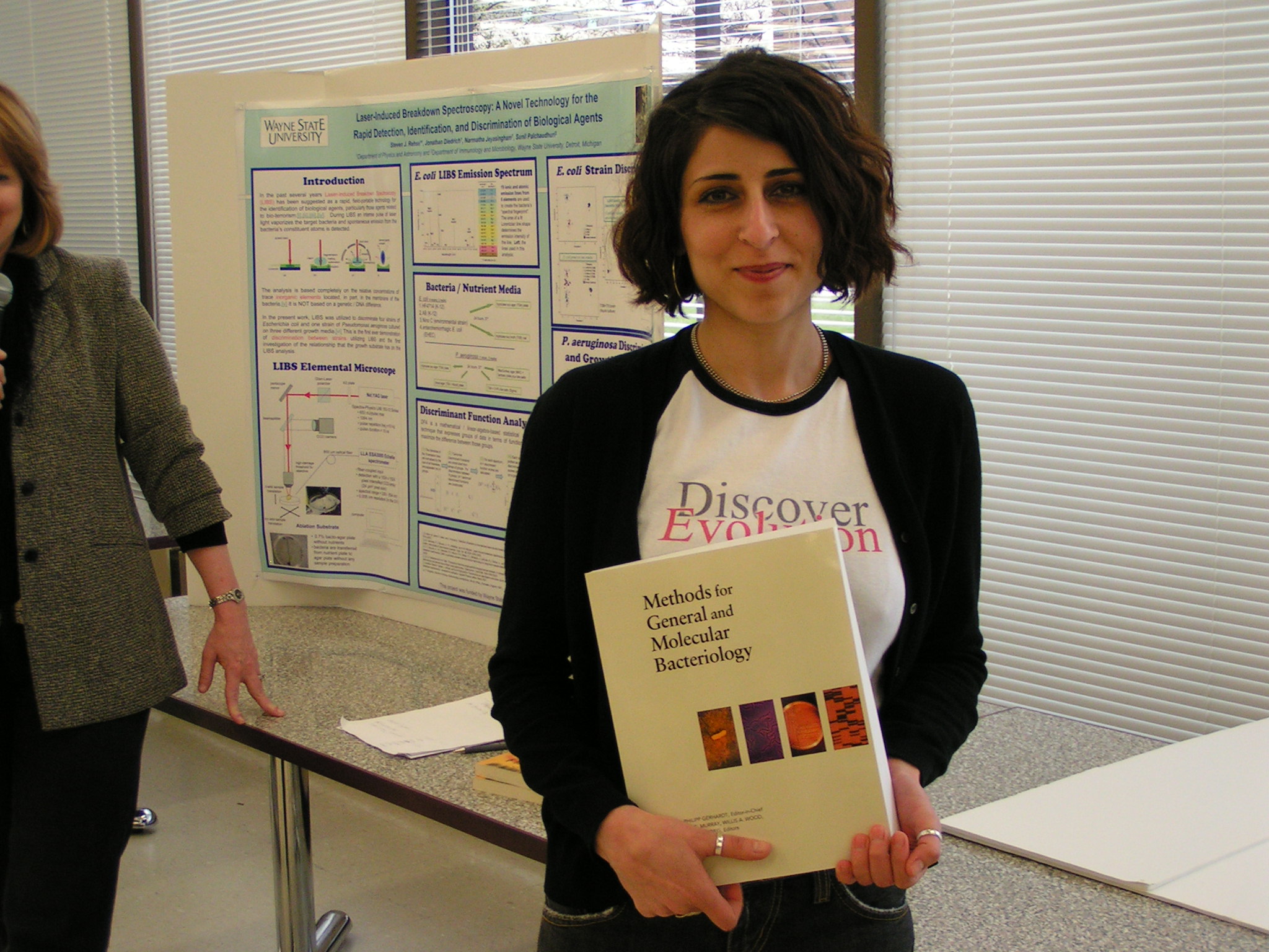
VISA, and MRSE. (received Feb 15) Laser-Induced Breakdown Spectroscopy: A Novel Technology
for the Laser-Induced Breakdown
Spectroscopy (LIBS) is an atomic spectroscopic technique that utilizes an intense
laser pulse to ablate a target and seed the constituent atoms into a high temperature
microplasma. Characteristic optical emission
from the plasma uniquely identifies the elemental composition of the vaporized target. Due to its inherent speed, accuracy, portability,
and simplicity, LIBS is currently being investigated as an early-warning technology for
the real-time detection and identification of harmful biological agents. We have utilized LIBS to rapidly identify and discriminate between
four strains of Escherichia coli, one strain of
environmental mold and one strain of Candida
albicans yeast. This was the first ever
demonstration of a rapid, efficient discrimination between different strains of a single
bacteria species based on the LIBS technology. The
effects of the bacteria growth environment were investigated by preparing samples of Pseudomonas aeruginosa on three different nutrient
media. Nearly identical spectra were obtained
from P. aeruginosa grown on TS agar and blood
agar plates, while the bacteria grown on a MacConkey plate exhibited easily
distinguishable differences indicating a chemical composition change, most likely in the
outer membrane of the bacteria. All samples of
P. aeruginosa were easily discriminated from
all E. coli strains. Isolation of Highly Beta-Hemolytic Bacteria for
Undergraduate Education Beta-hemolytic bacteria were isolated
from the environment as a pedagogical exercise. Microbes in aqueous soil suspension were
separated by streak plate inoculation on sheep blood agar. After incubation at 37oC,
among colonies of various sizes and pigmentation were some demonstrating a high degree of
beta-hemolysis. The appearance of selected colonies was recorded by a low power computer
microscope (Intel). Computer enhancement increased contrast and opportunity for learning.
The Gram stain revealed gram-positive bacilli and staining of aged culture with malachite
green showed spores. The hanging drop slide revealed motility. An antibiogram was prepared
by the antibiotic disc technique. Resistance to carbenicillin, sensitivity to kanamycin
and standard biochemical tests (glucose, citrate, mannitol, indole and catalase) provided
results consistent with those for Bacillus cereus. Identification
was supported by the Biolog automated redox-based system. The organism is recognized as a significant cause of
food poisoning. Isolation of beta-hemolytic bacteria from nature could acquaint students
with hemolysis and the characteristics of Bacillus
species of interest to public health. Antimicrobial Resistance in Escherichia
coli Isolates from Urinary Tract Infections in the Background:
The most common cause of urinary tract infections (UTIs) is Uropathogenic Escherichia coli (UPEC). Antimicrobial resistance
in UPEC isolates is highly varied and increasing. Objective:
To study UPEC strains in the
The Effect of Prostaglandin on Candida albicans-Dendritic
Cell Interactions
G. KUNDU* and M. NOVERR
Wayne State University, School of Medicine
-------------------------------------------------------------------------------------------------------
----------------------------------------------------------------------------------------------------------